From Classroom to Clinic to Patient: Anatomical Lung Models in Healthcare Practice
Anatomical lung models play an important role in medical education and practice, serving as invaluable tools for understanding respiratory anatomy, simulating disease progression, and perfecting clinical skills. For medical training providers and healthcare workers, the role of anatomical lung models is to bridge the gap between theoretical knowledge and practical application, offering a tangible representation of the complexities of the respiratory system.
Anatomical lung models can also be used effectively to explain complex health issues to patients.
Our bodies are incredibly complex and any tools that support a thorough understanding of how our organs work and interact are invaluable for the medical profession and patients alike. The role of anatomical lung models is certainly a fascinating one, so let’s just dig a little deeper.
Benefits of Using Lung Models in Medical Training
Enhancing Understanding
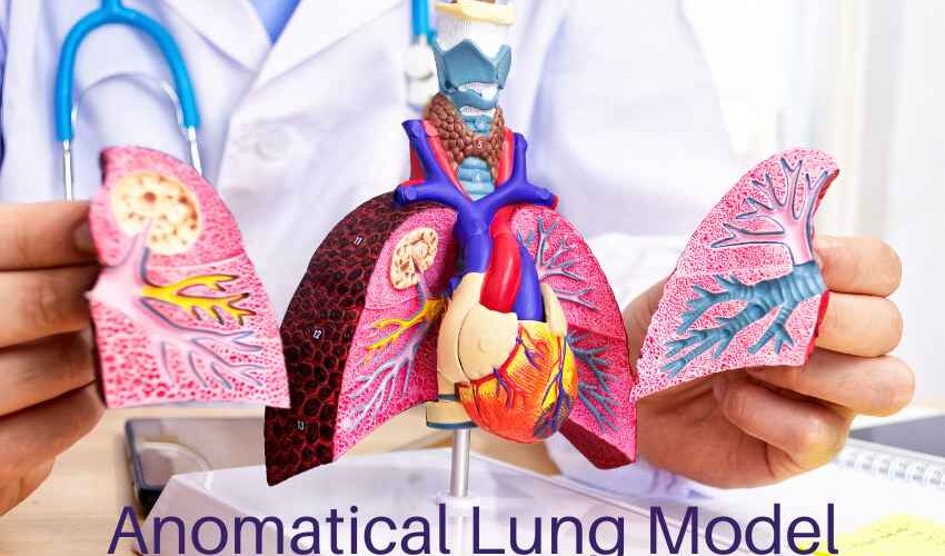
Anatomical lung models allow medical students and professionals to understand respiratory anatomy and physiology more effectively. Opposed to flat diagrams or textbooks, a full-sized lung model offers a tactile approach to exploring the complexities of this organ’s structure and function.
When students can see and touch the various parts of the lungs, from the bronchi to the alveoli, they develop a deeper understanding that enhances their clinical knowledge.
Observation of Different Disease Scenarios
These models also serve as dynamic teaching aids by simulating various respiratory diseases. You can find a lung model designed to showcase the appearance of different diseases such as chronic obstructive pulmonary disease (COPD), asthma, and lung cancer.
This simulation allows learners to develop diagnostic skills and treatment strategies without endangering real patients. For example, students can practice identifying characteristic features of different lung diseases, such as airway obstruction in COPD or the presence of nodules in lung cancer, and learn how to interpret diagnostic tests such as pulmonary function tests and chest X-rays.
This simulation-based approach not only improves clinical competence but also boosts confidence in managing real-life cases. By observing realistic disease scenarios in a controlled environment, students can practice making informed decisions and develop confidence in their abilities.
Skill Development
Practising procedures such as intubation and bronchoscopy on anatomical lung models help students become proficient before facing real patients. By familiarising themselves with the equipment and techniques in a simulated setting, students can refine their skills and build muscle memory, leading to smoother and safer procedures in clinical practice. This hands-on experience also allows them to troubleshoot potential challenges and develop strategies for managing complications.
Clinical Applications
Benefits to Patients
In clinical settings, anatomical lung models play a vital role in patient education and communication. When faced with a diagnosis or treatment plan related to respiratory health, patients often benefit from visual aids that make complex medical concepts easier to understand.
Anatomical lung models provide healthcare providers with a means to illustrate anatomy, disease processes, and treatment options in a clear and accessible manner, empowering patients to actively participate in their care decisions. By seeing a tangible representation of their condition, patients can better understand their prognosis and treatment options.
Planning Procedure & Interventions
Additionally, these models support pre-procedural planning, assisting medical teams in determining the best course of action for interventions. When getting ready for a bronchoscopy, biopsy, or surgical intervention, medical professionals can visualize anatomical landmarks, foresee possible obstacles, and determine the optimal course of action.
Types of Models
Basic lung anatomy models provide a basic understanding of respiratory structure and function, making them suitable for introductory training programs. Disease-specific models, such as those depicting COPD or lung cancer, offer targeted simulations for advanced clinical scenarios.
Then, there are functional models with respiratory mechanics simulation that provide a dynamic representation of lung physiology, allowing learners to explore concepts such as ventilation and gas exchange in a hands-on manner.
High-fidelity simulators with advanced features like real-time feedback and virtual reality integration offer an immersive training experience for advanced learners and seasoned professionals.
What Materials Are Lung Models Made of?
The most popular anatomical models are made of silicone as they are simple to shape and do not require any specialised tools. Silicone models, however, are not appropriate in all circumstances, especially when it comes to showing ultrasound-based operations. For example, the attenuation of ultrasound by silicone causes things to appear to be placed deeper than their real depth.
Anatomical models composed of synthetic and organic materials can be used for ultrasonography operations, making them a viable substitute for silicone models. Due to their reduced production costs, anatomical models constructed of organic materials, like gelatine and agar-agar gel, may be less expensive. However, they tend to dry up and disintegrate with time.
Anatomical models made of wood are also available, but they are primarily used as decor in doctors’ offices.
Synthetic anatomical models, including those made of polyvinyl alcohol (PVA) and polyvinyl chloride (PVC), are more durable than those constructed of organic materials. They can also be used repeatedly for simulations like bronchoscopy and intubation.
Anatomical models of foam are used to mimic the sponginess of the lungs to give trainees a better visual representation of these organs. And finally, there are 3D-printed models. They are not only realistic but also low-maintenance and simple to use. Their primary disadvantage is their high price.
How to Choose the Right Model
When selecting anatomical lung models for educational or clinical use, consider the following factors:
- Accuracy and Realism: Look for models that closely resemble the actual anatomy of the lungs for an effective learning experience. The accuracy of anatomical detail is crucial for providing learners with a realistic representation of respiratory anatomy and pathology.
- Durability and Materials: Consider the durability and materials used in construction. This is especially important for models intended for repeated use in clinical simulations. Models made from high-quality materials are more likely to withstand frequent handling and retain their anatomical fidelity over time.
- Compatibility with Educational Objectives: Evaluate the compatibility of models with specific educational objectives and curricula to ensure alignment with learning goals.
- Additional Features: Consider designs with additional features such as pathology simulation or interchangeable parts to enhance the versatility and utility of the learning experience. Models with interchangeable lungs or simulated pathologies allow learners to explore a wide range of clinical scenarios and develop diagnostic and treatment skills.
- Cost-effectiveness: When making your choice, take into account the models’ cost-effectiveness as well as any financial restrictions. High-fidelity simulators could be more expensive even though they might have more sophisticated features. Select the models that provide the most value for your clinical or educational environment by weighing the cost-benefit ratio.
To Sum Up
Using these medical simulation aids is a good way to promote respiratory education and improve the effectiveness of clinical practice. These aids offer a bridge between theory and practice, thus giving healthcare workers the knowledge they need to give their patients high-quality care.
As a patient, a better understanding of some of the learning materials and processes employed by the medial profession, such as the role of anatomical lung models, provides increased knowledge and confidence in the healthcare system.